
Introduction: The Precision of Ophthalmic Technology
Ophthalmology, the branch of medicine focused on eye health, demands a level of diagnostic and surgical precision unmatched in many other fields. The modern eye clinic must be equipped with sophisticated medical devices that allow practitioners to detect subtle pathologies, accurately measure ocular structures, and perform delicate surgical interventions. Investing in reliable, cutting-edge technology is paramount for delivering high-quality vision care and maintaining a competitive edge. This guide details the core equipment categories essential for any contemporary ophthalmology practice, emphasizing the crucial need for quality sourcing.

Pillar 1: Foundational Diagnostic Tools
Every comprehensive eye examination begins with a set of fundamental instruments designed to assess visual acuity and anterior segment health.
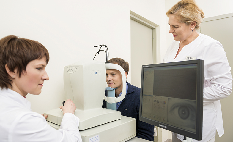
The Slit Lamp Biomicroscope
The Slit Lamp, or Biomicroscope, is the most crucial diagnostic tool in ophthalmology. It provides a highly magnified, three-dimensional view of the anterior segment of the eye (cornea, iris, lens) and, with specialized lenses, the posterior segment (retina and optic nerve). High-quality optics and stable mechanical movements are non-negotiable for accurate diagnosis of conditions ranging from cataracts to corneal ulcers.
Intraocular Pressure (IOP) Measurement
Measuring intraocular pressure is essential for screening and managing glaucoma. While various techniques exist, the gold standard is Applanation Tonometry (often mounted on the Slit Lamp). Reliable tonometers ensure accurate readings, which are vital for early intervention and preventing irreversible vision loss due to glaucoma.
Refractive Assessment: Autorefractors
Autorefractors quickly and objectively estimate a patient’s refractive error (myopia, hyperopia, astigmatism). These devices significantly streamline the examination process, providing a strong starting point for the subjective refraction performed using a Phoropter or trial lenses.
Pillar 2: Advanced Imaging and Structural Analysis
Modern ophthalmology relies heavily on cross-sectional and detailed structural imaging to manage complex diseases, particularly those affecting the retina and optic nerve.
Optical Coherence Tomography (OCT)
The Optical Coherence Tomography (OCT) machine is one of the most significant technological advancements in eye care. Using light waves, the OCT produces high-resolution, cross-sectional images of the retina and the optic nerve head, effectively allowing the clinician to view structures layer by layer. It is indispensable for diagnosing, monitoring, and managing macular degeneration, diabetic retinopathy, and glaucoma progression.
Fundus Cameras
Fundus Cameras capture detailed, high-resolution photographs of the back of the eye (fundus), which includes the retina, optic disc, and macula. These images serve as critical documentation for monitoring the progression of various retinal diseases and are invaluable for comparative analysis over time. The precision optics and complex analysis functions of these specialized imaging devices mirror the high standards of laboratory equipment like advanced Spectrophotometers and Analyzers required for scientific accuracy, standards which reputable general medical suppliers uphold.
Pillar 3: Surgical Excellence and Sterilization
For clinics offering surgical services, the quality of the operating equipment determines the outcome of the procedure.
The Surgical Operating Microscope
In procedures like cataract surgery, glaucoma surgery, and vitreoretinal surgery, an Operating Microscope is essential. Unlike standard laboratory microscopes, surgical microscopes must provide superior magnification, co-axial illumination, and freedom of movement, often integrating video recording capabilities. The requirement for exceptional optical clarity is a shared characteristic with the high-specification Microscopes supplied by general scientific vendors, though specialized ophthalmic units are tailored for surgical settings.
Phacoemulsification Machine
The Phacoemulsification Machine is the central device for modern cataract surgery. It uses ultrasonic energy to break up the cloudy lens, allowing the surgeon to remove it through a tiny incision. The machine’s fluidics and power management must be exceptionally reliable to ensure patient safety and surgical efficacy.
Infection Control: The Autoclave
The safety of any surgical environment depends on reliable sterilization. Specialized ophthalmic instruments, which are often delicate, must be meticulously cleaned and sterilized. A high-efficiency, certified Autoclave is mandatory for sterilizing surgical kits between cases. Sourcing such foundational equipment—whether it’s the Autoclave or Ultrasonic Cleaners for pre-cleaning—from quality-driven medical and laboratory suppliers, similar to the inventory offered by Eden International, is crucial for maintaining strict hygiene protocols.
Conclusion: A Holistic Investment in Quality
Equipping a successful ophthalmology clinic demands a holistic investment strategy. It requires not only the specialized ophthalmic units—like the OCT and Phaco machine—but also the highest quality supportive and foundational devices. The reliability of essential lab-grade equipment, such as Centrifuges (used for preparing biological eye treatments) and the core sterilization units, contributes directly to overall patient safety and clinical flow. By ensuring all medical devices meet rigorous standards, eye clinics solidify their reputation for providing advanced, safe, and effective vision care, positioning themselves for sustainable growth in the competitive medical market.
