

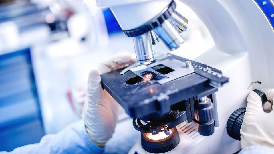
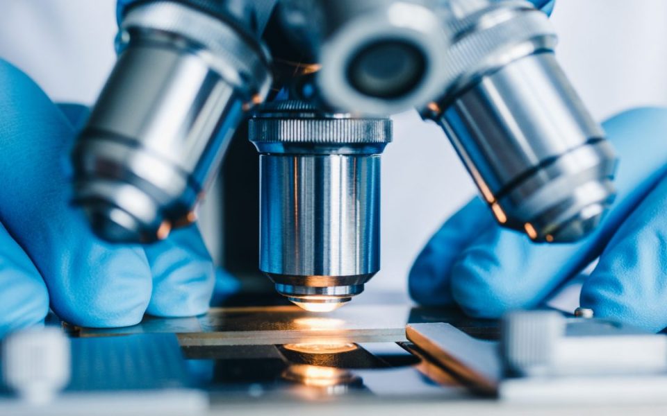
The quest to understand life at its most fundamental level—the cell—has historically been limited by the physical constraints of light. However, the last two decades have witnessed a revolution in optical microscopy, shattering the conventional diffraction limit and providing researchers with unprecedented views of cellular and molecular processes in situ. In 2026, advanced microscopy is no longer just a research tool; it is the cornerstone of modern cell biology, genomics, and regenerative medicine. From high-throughput screening in diagnostic laboratories, which require robust Compound Light Microscopes (like those supplied by medical distributors such as Eden International), to cutting-edge research facilities, the ability to visualize nanoscale dynamics is driving breakthroughs. This article explores the current state-of-the-art in advanced microscopy, focusing on its essential role in gene expression analysis and clinical diagnostics.
Shattering the Diffraction Limit: The Super-Resolution Revolution
Traditional fluorescence microscopy is restricted by Abbe’s diffraction limit, which prevents the resolution of structures smaller than approximately 200nm. The development of Super-Resolution Microscopy (SRM) techniques has entirely circumvented this barrier, providing the spatial resolution necessary to study molecular mechanisms, which are often in the 10nm to 50nm range.
Key SRM techniques include:
1.STED Microscopy (Stimulated Emission Depletion): This technique uses a second, red-shifted laser beam to de-excite fluorophores at the periphery of the illumination spot, effectively shrinking the Point Spread Function (PSF) and achieving resolutions down to 30nm axially and laterally.
2.SMLM (Single-Molecule Localization Microscopy): Encompassing techniques like STORM (Stochastic Optical Reconstruction Microscopy) and PALM (Photo-Activated Localization Microscopy), SMLM relies on mathematically reconstructing high-resolution images by temporally isolating and precisely localizing the positions of individual fluorophores. This is critical for molecular quantification and mapping protein-protein interactions within organelles.
3.Structured Illumination Microscopy (SIM): Though offering more modest resolution enhancement (typically a factor of two improvement over conventional widefield), SIM is valued for its high speed and low phototoxicity, making it an ideal choice for live-cell imaging experiments, such as tracking the dynamics of the cytoskeleton or the formation of invadopodia in cancer cells.
SRM’s ability to visualize individual protein complexes and nucleic acid structures is transformative for genetics, allowing researchers to track gene repair mechanisms, observe chromatin organization, and precisely locate specific mRNA molecules within the cytoplasm—all vital for understanding disease etiology.
Dynamic Imaging: Capturing Cellular Function in 4D
To truly understand cellular function, researchers must move beyond static snapshots to capture real-time processes. Techniques like Confocal Laser Scanning Microscopy (CLSM) and Multiphoton Microscopy are indispensable for this 3D and 4D (three dimensions plus time) imaging.
Confocal Microscopy achieves superior optical sectioning and contrast by utilizing a pinhole aperture positioned in the conjugate focal plane, which effectively rejects out-of-focus light and provides clean, high-contrast Z-stacks for 3D reconstruction. This capability is essential for studying complex tissues or organoids—the sophisticated 3D cell models used to mimic human organs.
For imaging deep within living tissues (in vivo or intravital imaging), Multiphoton Microscopy is the preferred method. It uses longer-wavelength infrared light for excitation, which scatters less and causes significantly less photodamage and photobleaching compared to conventional UV or visible light. This allows for deep penetration into thick samples, enabling studies of cellular migration within the native tissue microenvironment, such as monitoring immune cell trafficking or tumor growth kinetics. Furthermore, the integration of advanced techniques like FRET (Förster Resonance Energy Transfer) and FLIM (Fluorescence Lifetime Imaging Microscopy) with confocal systems allows for the measurement of molecular proximity and environmental changes (like Ca2+ concentration or pH) inside the cell, providing functional, not just structural, data
Integrating Microscopy for Precision Diagnostics and Research
The future of microscopy, as demonstrated by the availability of specialized laboratory tools and microscopes from suppliers like Eden International, lies in integration and automation.
The convergence of light and electron techniques, known as Correlative Light and Electron Microscopy (CLEM), allows researchers to link functional information (e.g., a fluorescently labeled protein’s activity seen via light microscopy) with the surrounding ultra-structural detail (e.g., the organelle context seen via Transmission Electron Microscopy – TEM or FIB-SEM). This holistic view is crucial for resolving the precise location of pathological processes, such as viral assembly or prion propagation, at the nanometer scale.
Furthermore, the rise of AI-Enhanced Imaging and Computational Image Analysis is transforming workflow efficiency. Machine learning algorithms are now employed for tasks such as automated feature recognition, cell segmentation, and high-throughput image analysis, allowing researchers to quickly process the massive datasets generated by high-content screening systems. In the clinical lab, advanced digital microscopy is central to modern cytopathology and biopsy analysis, enabling the precise identification of cellular abnormalities (like those seen in cancer or infectious diseases), thus enhancing the accuracy of diagnosis. The ability to image, analyze, and quantify is directly accelerating research in areas like neurodegenerative diseases and regenerative medicine.
Advanced microscopy, extending far beyond the limits of basic light imaging, serves as the critical enabler for molecular and cellular breakthroughs in the mid-2020s. Techniques like STED and SMLM now define the subcellular landscape, while confocal and multiphoton platforms reveal dynamic, 4D cellular communication within complex biological systems. By leveraging the power of super-resolution, live-cell imaging, and data integration (CLEM), researchers are gaining the essential knowledge needed to design gene therapies, optimize drug repurposing efforts, and enhance early disease diagnosis. As technology continues to integrate AI and push the boundaries of spatial and temporal resolution, the microscope remains the single most powerful scientific instrument, transforming the invisible world of the cell into actionable clinical and genetic insights.
